Nucleic Acids Life Sciences Grade 12 Notes and Study Guides. Nucleic acids are large biomolecules that are crucial in all cells and viruses. They are composed of nucleotides, which are the monomer components: a 5-carbon sugar, a phosphate group and a nitrogenous base. The two main classes of nucleic acids are deoxyribonucleic acid and ribonucleic acid
NUCLEIC ACIDS GRADE 12 NOTES – LIFE SCIENCES STUDY GUIDES
NUCLEIC ACIDS
LIFE SCIENCES
STUDY GUIDES AND NOTES
GRADE 12
CHAPTER 1:Nucleic acids
1.1 The structure of DNA and RNA
- Two kinds of nucleic acids are found in a cell, namely DNA and RNA.
- These two nucleic acids are made of building blocks (or monomers) called nucleotides.
- Figure 1.1 (right) shows what a nucleotide looks like.
Table 1.1 (below) shows the nitrogenous bases of DNA and RNA.

Table 1.1 Nitrogenous bases of DNA and RNA
- P – Phosphate group
- S – Deoxyribose or ribose sugar
- N – Nitrogenous base (adenine, thymine, guanine, cytosine or uracil)
Figure 1.1 below: A nucleotide

Figure 1.2 below shows the structure of DNA and RNA. Study the diagrams

in Figure 1.2, and then read the information in the boxes below the diagrams to find out how to tell a DNA molecule from an RNA molecule.
 Figure 1.2 The structure of DNA and RNA
| How to recognise a DNA molecule | How to recognise an RNA molecule |
|
|
|
|
| |
|
1.2 Differences between DNA and RNA
Table 1.2 below summarises the differences between DNA and RNA molecules.
| DNA | RNA |
| 1. Double-stranded molecule | 1. Single-stranded molecule |
| 2. Contains deoxyribose (sugar) | 2. Contains ribose (sugar) |
| 3. Contains the nitrogenous base, thymine | 3. Contains the nitrogenous base, uracil |
Table 1.2 The differences between DNA and RNA
1.3 DNA replication
DNA replication takes place at interphase before mitosis or meiosis begins. DNA replication is the process during which a DNA molecule makes an exact copy (replica) of itself. This is shown in Figure 1.3 below.

Figure 1.3 DNA replication
- The double helix CG unwinds.
- Weak hydrogen bonds between nitrogenous bases break and two DNA strands unzip (separate).
- Each original DNA strand serves as a template on which its complement is built.
- Free nucleotides build a DNA strand onto each of the original two DNA strands by attaching to their complementary nitrogenous bases (A to T and C to G).
- This results in two identical DNA molecules. Each molecule consists of one original strand and one new strand.
Significance of DNA replication
DNA replication is important because it:
- Doubles the genetic material so it can be shared between the resulting daughter cells during cell division.
- Results in the formation of identical daughter cells during mitosis.
1.4 DNA profiling
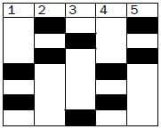
Every person except identical twins has her/his own unique DNA profile. It can be described as an arrangement of black bars representing DNA fragments of the person.
It is used to:
- Identify criminals
- Identify dead bodies
- Identify relatives
- Identify paternity
Activity 1
- A DNA molecule contains 600 nitrogen bases. If 20% of this is adenine, determine the number of each nitrogen base in the DNA molecule. (3)
- Figure 1.4 (below) represents part of a nucleic acid molecule. Study the diagram and answer the questions that follow.
2.1 Identify the nucleic acid shown in Figure 1.4. (1)
2.2 Label the following: (drawing below)- Part 1 (1)
- Part 2 (1)
- The nitrogenous bases 4, 5 and 6 (3)
2.3 What is the collective name for the parts numbered 1, 2 and 3?(1)
- Questions 3.1 and 3.2 are based on Figure 1.5 (below). This is a diagrammatic representation of a part of two different nucleic acid molecules found in the cells of organisms during a stage in the process of protein synthesis.
3.1 Name the molecules 1 and 2. (2)
3.2 Give a reason for your answer in question 3.1. (2) - The result of profiling various DNA samples in a criminal investigation is shown below
 (Key below}
4.1 Was suspect X or suspect Y involved in the crime? (1)
4.2 Does the DNA of the suspect (from answer 4.1) match the first or second sample? (2) [17]
| Key for question 4: | Drawings for Q2 and Q3 above |
|  |
Answers to activity 1
- 20% adenine = 20% thymine✔ 30% cytosine✔= 30% guanine✔
20 × 600 = 120A = 120T 30 × 600 = 180C = 180G (3)
100 100 - 2.1 DNA✔
2.2- Phosphate✔ group (1)
- Deoxyribose✔sugar (1)
- 4 – adenine (A)✔
6 – thymine✔
5 – guanine (G)✔ (3)
2.3 Nucleotide✔ (1)
- 3.1 1 – DNA 2 – mRNA/RNA✔ (2)
3.2 DNA contains the nitrogenous base thymine (T).✔
RNA contains the nitrogenous base uracil (U).✔ (2) - 4.1 Suspect X was involved. ✔(1)
4.2 The DNA of suspect X matches with the second sample. ✔✔ (2) [17]
1.5 Protein synthesis
Protein synthesis is the process by which proteins are made in each cell of an organism to form enzymes, hormones and new structures for cells.

Figure 1.6 The process of protein synthesis
There are two main processes involved in protein synthesis, namely transcription and translation. They are labelled as A and B in Figure 1.6 above.
- Note that the numbers on the diagram correspond with the description below.
A Transcription (takes place in the nucleus)
- DNA unwinds and splits.
- One DNA strand acts as a template for forming mRNA.
- Free nucleotides arrange to form mRNA according to the DNA template. This process is called transcription.
- The mRNA leaves the nucleus through the nuclear pores. Stage
B now takes place when mRNA in the cytoplasm attaches to the ribosome.
B Translation (takes place in the cytoplasm on the ribosome)
5. Each tRNA brings a specific amino acid to the mRNA. This is called translation.
6. The amino acids are linked together to form a particular protein.
The diagram shown in Figure 1.6 (on page 5) may appear in exam questions in different ways. Do not let the different representations confuse you. Just try to identify the
following components by looking for the features listed here:
- DNA – double-stranded; look for presence of thymine; found in nucleus only.
- Nuclear membrane – has nuclear pores through which mRNA moves.
- mRNA – single-stranded; look for presence of uracil; contains a triplet of bases (codon) found in nucleus and cytoplasm.
- Ribosome – usually mRNA attached to it.
- tRNA – contains a triplet of bases (anticodon); look for attached amino acid.
Activity 2
Question 1
Study Figure 1.7 (below), which shows the process of protein synthesis, and answer the questions.

Figure 1.7 Protein synthesis
1.1 Label structures A, B and D. (3)
1.2 State ONE function of molecule D. (1)
1.3 Which stage of protein synthesis takes place at F? (1)
1.4 Identify organelle C. (1)
1.5 Name and describe the stage of protein synthesis that takes place at organelle C. (7)
1.6 Write down the codon of anticodon E from top to bottom. (1)
1.7 Name the type of bond (labelled G) between the amino acids. (1) [15]
Answers to question 1
1.1
A – Nuclear membrane✔
B – mRNA✔
D – DNA✔ (3)
1.2 Carrying hereditary characteristics from parents to their offspring✔ OR Controls the synthesis (manufacturing) of proteins✔ (1)
1.3 Transcription✔ (1)
1.4 Ribosome✔ (1)
1.5 Translation✔
- The mRNA strand from the nucleus becomes attached✔to a ribosome with its codons exposed
- each tRNA molecule carrying a specific amino acid✔
- according to its anticodon✔
- matches up with/complements the codon of the mRNA✔
- so that the amino acids are placed in the correct sequence✔
- adjacent amino acids are linked✔
- to form a protein✔ (7)
1.6 CAC✔ (the anticodon is GUG, so the complementary codon is CAC) (1)
1.7 Peptide Bond (1) [15]
Question 2
Table 1.3 below shows the DNA base triplets that code for different amino acids.
| Amino acid | Base triplet in DNA template |
| Leu (leucine) | GAA |
| His (histidine) | GTA |
| Lys (lysine) | TTT |
| Pro (proline) | GGG |
| Ala (alanine) | CGA |
| Trp (tryptophan) | ACC |
| Phe (phenylalanine) | AAA |
| Gly (glycine) | CCT |
Table 1.3 Different amino acids and their DNA base triplets
The following is a part of a sequence of amino acids that forms a particular protein molecule:
| Ala | His | Trp | Leu | Lys |
2.1 Name the process by which mRNA is formed from a DNA template. (1)
2.2 How many mRNA codons would be involved in forming the portion of protein shown above? (1)
2.3 Write down the sequence of the first three mRNA codons (from left to right) for this portion of the protein. (3) [5]
Answers to question 2
2.1 Transcription✔ (1)
2.2 5✔ (1)
2.3 GCU✔- CAU✔- UGG✔ (3) [5]

 (Key below}